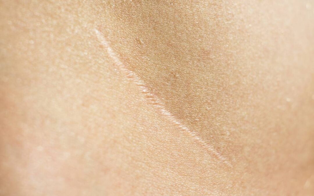
The burden of wounds is hefty across the United States: in 2018, a retrospective study of Medicare beneficiaries revealed that approximately 8.2 million beneficiaries suffered from infectious and non-infectious wounds in 2014 (1). Wound-associated scars – especially those resulting from chemical and thermal burns – can result in disfigurement, disability, and poorer quality of life, and treatments to minimize such side effects are costly, with the financial burden of scar treatments estimated to reach approximately $32 billion worldwide by 2027 (2). Scars can also occur as a result of surgery. Elucidating the underlying mechanism of scarring is therefore critical to ensuring optimized clinical interventions in order to minimize complications (3). The underlying molecular and cellular pathways of these processes are not yet completely understood but are slowly being unraveled.
In normal, healthy tissues, both fibroblasts and myofibroblasts secrete remodeling enzymes and metalloproteinases that enable the dynamic remodeling of the extracellular matrix in the context of a dynamic, bidirectional dialogue with their local microenvironment. In tissue repair in response to a wound, myofibroblasts enable the formation of granulation tissue via growth factor-β1-induced myofibroblastic differentiation (4). This proceeds alongside precisely coordinated myofibroblast apoptosis and extracellular matrix remodeling, key to healthy scar formation (5).
At a molecular level, the underlying mechanisms of this cellular dance are complex. First, the mechanical forces that cells are subjected to activate mechanical signaling to control myofibroblast phenotype. Specifically, a recent study found that during wound healing, the Engrailed-1 (En1) gene is expressed in En1-lineage–negative fibroblasts of the deep dermal layer as a result of increased tensile forces (6). When the extracellular matrix stiffens, tensile forces are cellularly transmitted by allosteric modulation of the integrin cytoplasmic domain. This triggers a signal transduction pathway leading to the translocation of Yes-associated protein (YAP) to the nucleus, where it triggers the conversion of En1-lineage–negative fibroblasts to En1-lineage–positive fibroblasts. This activates a fibrotic response, generating more traction and collagen deposition. In En1 knockout mice or mice in which YAP function is genetically or pharmacologically disrupted via the small, Food and Drug Administration pre-approved molecule verteporfin, wounds heal more slowly and do not scar.
Second, innervation and the presence of local inflammation are equally involved in both skin repair processes and the differentiation of myofibroblasts, including in the context of hypertrophic scars (4). Normal innervation and the associated secretion of key neuropeptides are critical to maintaining skin homeostatic mechanisms and healthy wound healing; however, the precise roles of autonomic and sensory innervation remain to be deciphered (7).
Results elucidating the cellular and molecular mechanism of scarring may advance the treatment of certain fibrotic disorders. For example, verteporfin was described as an inhibitor of fibrosis in patients with persistent cholestasis, after which it was found to also prevent fibrosis in several human organs, including the lung. However, preclinical studies are warranted to confirm these findings in more human-like clinical models. In addition, verteporfin is not expected to block the formation of keloids, which arise primarily from excess cell proliferation rather than extracellular matrix production.
In view of the burden of human wounds, including those associated with international conflict and forest fires, a consortium of more than 75 national institutions, including the Department of Defense and National Institutes of Health, has galvanized research on scarless healing and regenerative healing. Ongoing research will continue to allow us to better dissect the precise molecular and biological mechanism of wound scarring.
References
- Nussbaum SR, Carter MJ, Fife CE, DaVanzo J, Haught R, Nusgart M, et al. An Economic Evaluation of the Impact, Cost, and Medicare Policy Implications of Chronic Nonhealing Wounds. Value Heal. 2018;
- Sen CK. Human Wound and Its Burden: Updated 2020 Compendium of Estimates. Advances in Wound Care. 2021.
- Clark RAF. To Scar or Not to Scar. https://doi.org/101056/NEJMcibr2107204. 2021 Jul 28;385(5):469–71.
- Darby IA, Desmoulière A. Scar Formation: Cellular Mechanisms. In: Textbook on Scar Management. 2020.
- Hinz B, Lagares D. Evasion of apoptosis by myofibroblasts: a hallmark of fibrotic diseases. Nature Reviews Rheumatology. 2020.
- Mascharak S, des Jardins-Park HE, Davitt MF, Griffin M, Borrelli MR, Moore AL, et al. Preventing Engrailed-1 activation in fibroblasts yields wound regeneration without scarring. Science (80- ). 2021;
- Laverdet B, Danigo A, Girard D, Magy L, Demiot C, Desmoulière A. Skin innervation: Important roles during normal and pathological cutaneous repair. Histology and Histopathology. 2015.
- Rinkevich Y, Walmsley GG, Hu MS, Maan ZN, Newman AM, Drukker M, et al. Identification and isolation of a dermal lineage with intrinsic fibrogenic potential. Science (80- ). 2015;


Recent Comments