
For Informational Purposes Only
Imaging Guidance for Nerve Blocks: Analyzing Ultrasound Images
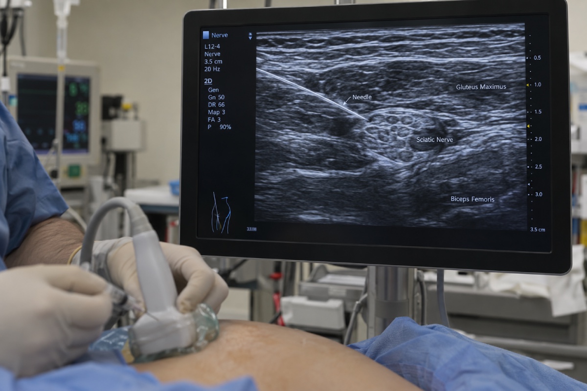
Ultrasound is an integral component of regional anesthesia, allowing anesthesiologists to visualize nerves, surrounding anatomy, needle advancement, and local anesthetic spread in real time. Compared with landmark- and nerve-stimulation techniques, ultrasound guidance can improve procedural precision and reduce the incidence of vascular puncture and other complications. However, the clinical value of ultrasound depends not simply on placing a probe over the target but on accurately interpreting the image and translating two-dimensional anatomy into safe three-dimensional needle movement. For the anesthesiologist, skill in analyzing ultrasound images for nerve block guidance is increasingly important.
A high-quality ultrasound examination begins with appropriate probe selection, patient positioning, and optimization of imaging parameters. High-frequency linear transducers provide excellent resolution for superficial structures, whereas lower-frequency curvilinear probes are necessary for deep targets. Depth, gain, focus, and dynamic range should be adjusted to obtain the clearest possible distinction between neural structures and surrounding tissues. The anesthesiologist should first identify consistent anatomic landmarks before attempting needle advancement. Arteries, veins, muscles, fascia, bones, tendons, and pleura provide orientation and help establish the relationship between the intended nerve and structures that must be avoided.
Peripheral nerves have characteristic but not identical sonographic appearances. In cross-section, many nerves demonstrate a hypoechoic fascicular pattern surrounded by more echogenic connective tissue. Their appearance changes according to anatomical location, patient age, tissue depth, and the orientation of the transducer. Consequently, a nerve should not be identified solely on the basis of a single visual characteristic. Dynamic scanning, following the structure proximally and distally, and correlating the ultrasound image with known anatomy are important steps when searching for the target nerve for the nerve block. This is particularly important when nerves are small, deeply situated, or surrounded by structures with similar echogenicity.
Needle visualization is another critical component of ultrasound-guided regional anesthesia. In-plane needle insertion enables the visualization of a larger portion of the needle shaft and tip, whereas out-of-plane techniques may provide only a cross-sectional view of the needle. Regardless of the approach, identification of the needle tip is essential. Advancing a needle when only the shaft is visible can result in unintended deep penetration and injury to nerves, vessels, pleura, or other structures. Small adjustments in probe position, needle angle, and beam alignment can substantially improve needle visibility. Hydrolocalization with small volumes of fluid may also help confirm the needle tip when visualization is difficult.
Assessment of local anesthetic spread provides an additional safety mechanism. Rather than injecting a predetermined volume without observing its distribution, the anesthesiologist can use ultrasound to determine whether the injectate is spreading around the intended neural structure. Circumferential or appropriately targeted spread may confirm correct needle positioning, whereas intraneural expansion, significant vascular injection, or unexpected tissue-plane spread should prompt immediate reassessment. Injection should always remain low pressure and incremental, with continuous attention to patient symptoms and signs of local anesthetic systemic toxicity.
Ultrasound imaging also has limitations. Poor image quality may result from obesity, edema, calcification, deep anatomy, or inadequate probe positioning. Acoustic shadowing from bone and anisotropy can obscure important structures. Furthermore, ultrasound provides a two-dimensional representation of three-dimensional anatomy, and apparent proximity on the screen does not necessarily correspond to actual spatial relationships. Anesthesiologists analyzing the ultrasound image therefore must have both image recognition skills and an understanding of anatomy, sonoanatomy, needle mechanics, and potential complications when performing image-guided nerve block.
Ultrasound is a dynamic clinical examination rather than simply a needle-navigation device. A systematic sequence of identifying anatomy, optimizing the image, confirming the target, locating the needle tip, observing injectate spread, and reassessing the final position can improve procedural safety. As regional anesthesia continues to evolve, proficiency in ultrasound image interpretation will remain central to achieving reliable nerve blocks while minimizing complications.
References
- Neal JM, Barrington MJ, Fettiplace MR, et al. The third American Society of Regional Anesthesia and Pain Medicine practice advisory on local anesthetic systemic toxicity: executive summary 2023. Reg Anesth Pain Med. 2023;48(3):113-123. DOI: https://doi.org/10.1097/aap.0000000000000720
- Barrington MJ, Kluger R. Ultrasound guidance reduces the risk of local anesthetic systemic toxicity following peripheral nerve blockade. Reg Anesth Pain Med. 2013;38(4):289-299. DOI: https://doi.org/10.1097/aap.0b013e318292669b
- Perlas A, Chan VWS, Beattie S. Anesthesia and analgesia in the ambulatory setting: ultrasound-guided regional anesthesia. Anesthesiol Clin. 2010;28(2):315-330.
- Sites BD, Spence BC, Gallagher J, et al. Characterizing novice behavior associated with learning ultrasound-guided peripheral regional anesthesia. Reg Anesth Pain Med. 2007;32(2):107-115. DOI: https://doi.org/10.1016/j.rapm.2006.11.006
- Albrecht E, Chin KJ. Advances in regional anesthesia and acute pain management: ultrasound-guided regional anesthesia. Anesthesiology. 2020;132(5):1082-1098. DOI: https://doi.org/10.1111/anae.14868